Joint · Lower limb
Ankle joint anatomy: bones, ligaments and muscles
The ankle joint links the tibia and fibula to the talus. Learn the bones of the ankle, its medial and lateral ligaments and the muscles that move the foot.
The ankle joint, or talocrural joint, is the synovial hinge joint where the lower ends of the tibia and fibula grip the talus. It only lifts the foot up (dorsiflexion) and points it down (plantar flexion); the muscles in front of the shin lift it, the calf muscles point it, and ligaments on each side hold the talus in place.
Which bones form the ankle joint?
Three bones meet at the ankle joint:
- The tibia, the shin bone. Its lower end carries the body's weight onto the talus, and its medial malleolus is the bump on the inner side of the ankle.
- The fibula, the thin outer bone of the leg. Its lower end, the lateral malleolus, is the bump on the outer side of the ankle.
- The talus, the top bone of the foot. Its rounded, pulley-shaped upper surface, the trochlea, fits between the two malleoli.
Picture a mortise and tenon, the woodworking joint: the tibia and the two malleoli make the socket, and the talus is the piece that fits into it. The tibia sits on the top of the talus and carries the load, while each malleolus covers one side. Held in this socket, the talus can rock forward and back but cannot shift sideways.
Are the tarsal bones "ankle bones" too?
Often, yes, in everyday speech. Strictly, only the tibia, the fibula and the talus form the joint, but "the ankle bones" often also means the seven tarsal bones at the back of the foot: the talus, calcaneus, navicular, cuboid and the three cuneiforms. When you stand, your weight goes down the tibia into the talus and on to the heel bone under it. The bones of the foot table lists all 26 bones of the foot and the muscles attached to each.
What movements happen at the ankle?
| Movement | Where it happens | What it does |
|---|---|---|
| Dorsiflexion | Ankle (talocrural) joint | Lifts the top of the foot toward the shin |
| Plantar flexion | Ankle (talocrural) joint | Points the foot and toes down, lifts the heel |
| Inversion | Subtalar joint and other joints between the tarsal bones | Turns the sole inward |
| Eversion | Subtalar joint and other joints between the tarsal bones | Turns the sole outward |
The ankle joint is a hinge with one axis, so it cannot turn the sole. That job belongs to the joints below the talus: first the subtalar joint between the talus and the calcaneus, then the talonavicular and calcaneocuboid joints further forward. Each of them glides only a little.
Which ligaments hold the ankle together?
The front and back of the joint are left free for the foot's up-and-down swing. The strong ligaments are on the inner and outer sides, each starting on a malleolus:
| Side | Ligament | Runs between | Resists |
|---|---|---|---|
| Medial (inner) | Deltoid ligament, a broad fan | Medial malleolus of the tibia and the tarsal bones below it, mainly the talus and calcaneus | Too much eversion |
| Lateral (outer) | Anterior talofibular ligament | Lateral malleolus of the fibula and the talus, in front | Too much inversion |
| Lateral (outer) | Posterior talofibular ligament | Lateral malleolus of the fibula and the talus, behind | Too much inversion |
| Lateral (outer) | Calcaneofibular ligament | Lateral malleolus of the fibula and the calcaneus | Too much inversion |
Which muscles move the ankle?
No muscle attaches to the talus. The muscles of the leg cross the ankle and end on the bones of the foot, so they move the foot, and the talus moves with it under the tibia and fibula.
| Movement | Main muscles | Compartment and nerve |
|---|---|---|
| Dorsiflexion | Tibialis anterior, extensor digitorum longus, extensor hallucis longus | Anterior compartment, deep fibular nerve |
| Plantar flexion | Gastrocnemius and soleus through the calcaneal (Achilles) tendon, helped by the tibialis posterior, flexor digitorum longus, flexor hallucis longus and the fibularis muscles | Posterior compartment, tibial nerve (fibularis muscles: lateral compartment) |
| Inversion | Tibialis anterior, tibialis posterior | Anterior compartment, deep fibular nerve (tibialis anterior); posterior compartment, tibial nerve (tibialis posterior) |
| Eversion | Fibularis longus, fibularis brevis | Lateral compartment, superficial fibular nerve |
The tendons of the dorsiflexors run under the superior and inferior extensor retinacula, two straps across the front of the ankle that stop them lifting away from the joint.
How to study the ankle joint in 3D
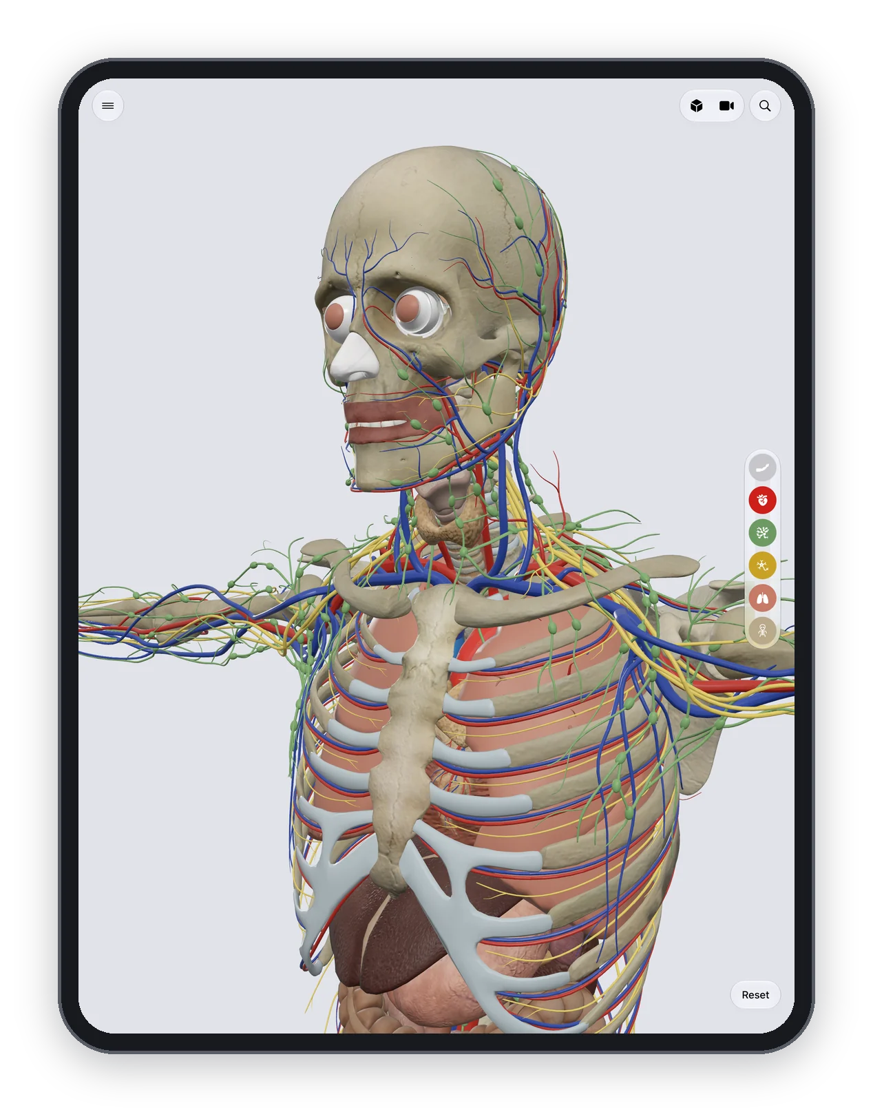
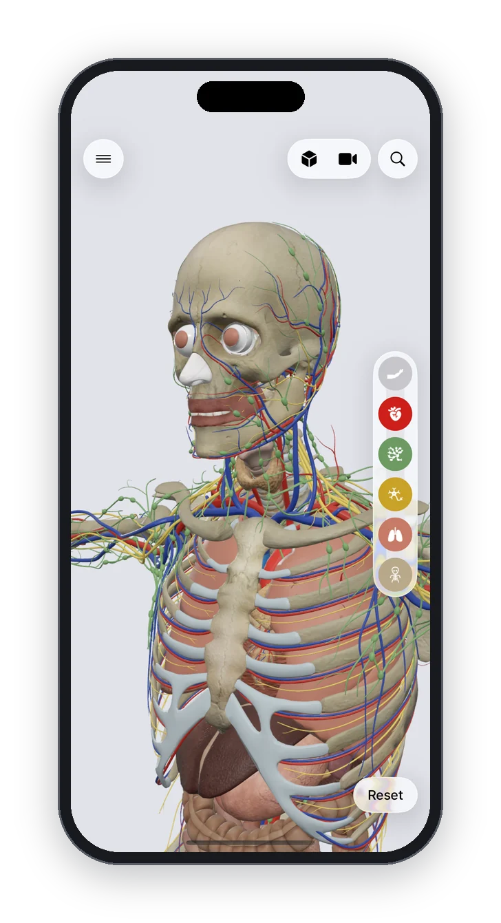
Anatomy Atlas 3D is a free 3D human anatomy atlas for iPad and iPhone: 2,000 structures, no account, no ads, works offline, English and Latin names, and a tap-the-structure quiz. It shows the bones of the ankle and foot and the muscles of the leg, but not the ankle ligaments or the retinacula, so use it for the shape of the joint and the path of each muscle. The malleoli are parts of the tibia and fibula, not separate labels:
- Peel away the muscle layer and look at the ankle from the front. Find the lower end of the tibia, the fibula on the outside and the talus between them.
- Turn the foot to see the medial malleolus on the inner side and the lateral malleolus on the outer side, then follow the talus down to the calcaneus.
- Bring the muscles back and tap the tibialis anterior, then the gastrocnemius and the soleus, to read where each one ends on the foot.
- Test yourself with the leg and foot quiz or the lower limb muscles quiz, and find every lower limb page on the lower limb hub.
The knee joint and the elbow joint are the other big hinges of the body, with their own pairs of collateral ligaments.
Frequently asked questions
What type of joint is the ankle?
A synovial hinge joint with one axis. It allows dorsiflexion and plantar flexion only; turning the sole in and out happens at the subtalar joint below it.
What 3 bones make up the ankle joint?
The tibia, the fibula and the talus. The lower ends of the tibia and fibula form a slot that holds the top of the talus.
What are the bumps on each side of the ankle?
The medial malleolus on the inner side is the lower end of the tibia. The lateral malleolus on the outer side is the lower end of the fibula.
What are the ligaments of the ankle?
The deltoid ligament on the medial side, and three ligaments on the lateral side: the anterior talofibular, the posterior talofibular and the calcaneofibular ligaments.
Which muscles point the foot down?
The gastrocnemius and the soleus, through the calcaneal (Achilles) tendon on the heel, helped by the deep calf muscles and the fibularis muscles.
Is the subtalar joint part of the ankle joint?
No. The subtalar joint lies below the ankle joint, between the talus and the calcaneus. It turns the sole in and out, while the ankle joint moves the foot up and down.
