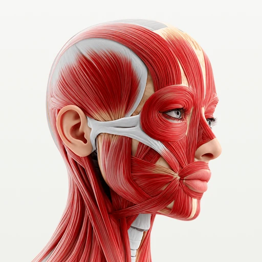
Joint · Head and neck
Atlanto-axial joint: how the atlas and axis move
The atlas turns around the dens of the axis at three atlanto-axial joints, the "no" movement. Learn the three joints, the nodding joint above and the ligaments.
The atlanto-axial joint is where the atlas (C1) turns on the axis (C2): the ring of the atlas, carrying the skull, rotates around the dens, the peg of bone that rises from the axis. It is the joint of the "no" movement. Nodding "yes" happens one level higher, at the atlanto-occipital joints between the skull and the atlas.
How do the atlas and axis move?
Their main movement is rotation. When you turn your head to look over your shoulder, the skull and the atlas turn together as one piece around the dens of the axis, while the axis stays in place below them. Much of the turning of the neck happens at this one level; the joints between the lower cervical vertebrae add the rest. The atlas can also tip forward and back a little on the dens.
The two levels at the top of the neck split the work between them:
| Joint | Bones | Type | Main movement |
|---|---|---|---|
| Atlanto-occipital joints (left and right) | Occipital condyles of the skull and the superior facets of the atlas | Synovial, condylar | Nodding "yes" (flexion and extension of the head), a little side bending |
| Median atlanto-axial joint | Dens of the axis and the anterior arch of the atlas, closed behind by the transverse ligament | Synovial, pivot | Turning "no" (rotation) |
| Lateral atlanto-axial joints (left and right) | Inferior facets of the atlas and superior facets of the axis | Synovial, plane | Glide during rotation and carry the weight of the head |
What are the three atlanto-axial joints?
The atlanto-axial joint is really three joints that work together:
- The median atlanto-axial joint, in the midline. The front of the dens touches a small facet on the back of the anterior arch of the atlas. Behind the dens, the transverse ligament of the atlas closes the ring, so the dens turns inside a loop that is half bone and half ligament. This is the classic example of a pivot joint.
- The left and right lateral atlanto-axial joints. Each lateral mass of the atlas sits on one of the broad upper facets of the axis, which slope gently down toward the sides. The facets glide over each other as the atlas turns, each with its own loose capsule.
There is no intervertebral disc between the atlas and the axis, and none between the skull and the atlas. The first disc of the spine sits between C2 and C3, which is one reason the top of the neck turns so much more freely than the levels below it.
What happens at the atlanto-occipital joint?
The two occipital condyles on the base of the skull sit in the cupped upper facets of the atlas. Each condyle sits in its cup like the curved base of a rocking chair: tipping the skull forward and back over the atlas is the nod. The joint also lets the head tilt a little to the side, but it barely turns, so turning the head is left to the joints below.
Which ligaments hold the atlas, axis and skull together?
| Ligament | Runs between | What it does |
|---|---|---|
| Transverse ligament of the atlas | The inner sides of the two lateral masses of the atlas, behind the dens | Holds the dens against the anterior arch; the main stabilizer of the atlanto-axial joint |
| Cruciate (cruciform) ligament of the atlas | The transverse ligament plus a band up to the skull and a band down to the axis | Makes a cross behind the dens |
| Alar ligaments (left and right) | The sides of the tip of the dens and the inner sides of the occipital condyles | Limit how far the head can turn |
| Apical ligament of the dens | The tip of the dens and the front edge of the foramen magnum | A thin cord in the midline |
| Tectorial membrane | The back of the body of the axis and the inside of the base of the skull | Covers the cruciate and alar ligaments from behind, on the side of the spinal canal; it continues the posterior longitudinal ligament of the spine |
| Anterior and posterior atlanto-occipital membranes | The arches of the atlas and the edges of the foramen magnum | Close the gaps in front of and behind the atlanto-occipital joints |
Which muscles turn and nod the head?
No single muscle runs only from the atlas to the axis to rotate it. The head is turned by muscles of the neck that act on the skull and the upper vertebrae, often working with the same muscle on the other side:
| Movement | Main muscles | How they act |
|---|---|---|
| Turning the head (rotation) | Sternocleidomastoid | One side turns the face to the opposite side |
| Turning the head (rotation) | Splenius capitis and splenius cervicis | One side turns the head and neck to the same side |
| Turning the head (rotation) | Obliquus capitis inferior and rectus capitis posterior major, two suboccipital muscles | Turn the atlas and the skull to the same side; the obliquus capitis inferior runs from the spinous process of the axis to the transverse process of the atlas |
| Nodding forward (flexion) | Both sternocleidomastoids, with the deep muscles in front of the spine | Bend the head and neck forward |
| Lifting the head back (extension) | Both splenius capitis, the upper trapezius, and the rectus capitis posterior major and minor and obliquus capitis superior (suboccipital muscles) | Tip the head back on the atlas |
The suboccipital muscles lie deep under the base of the skull and are supplied by the suboccipital nerve, the posterior branch of the first cervical nerve. The sternocleidomastoid is supplied by the accessory nerve (cranial nerve XI).
How to study the atlas and axis joints in 3D
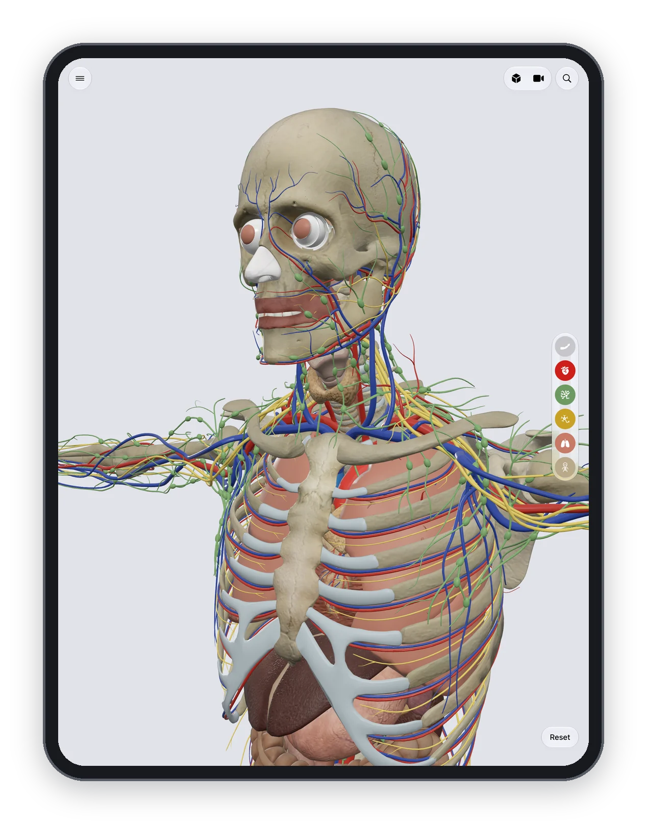
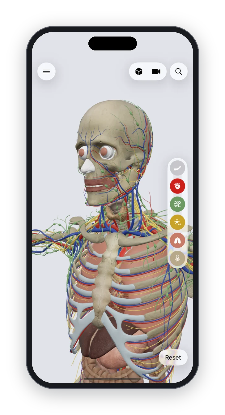
Anatomy Atlas 3D is a free 3D human anatomy atlas for iPad and iPhone: 2,000 structures, no account, no ads, works offline, English and Latin names, and a tap-the-structure quiz. It shows the skull, the atlas, the axis and the other cervical vertebrae, and the large neck muscles such as the sternocleidomastoid and the splenius cervicis. It does not show the ligaments of this region or the small suboccipital muscles, so use it for the shape of the bones and the way they fit:
- Peel away the muscles of the neck and tap the "Atlas (C1 vertebra)", then the "Axis (C2 vertebra)" just below it.
- Look at the two from above: find the dens of the axis sitting inside the front of the ring of the atlas.
- Turn the model to the side to see the lateral masses of the atlas resting on the axis, and the skull resting on the atlas.
- Bring the muscles back and tap the sternocleidomastoid on the side of the neck, then test yourself with the neck quiz.
The cervical vertebrae page compares C1 and C2 with the five vertebrae below them, the bones of the spine table lists every vertebra, and the head and neck hub gathers every page of the region. The body's other classic pivot is the proximal radioulnar joint, inside the capsule of the elbow joint, where the head of the radius turns in a ring of ligament.
Frequently asked questions
What type of joint is the atlanto-axial joint?
Synovial. The middle joint, between the dens and the atlas, is a pivot joint that allows rotation only; the two lateral joints are flat plane joints that glide as the atlas turns.
Which joint lets you shake your head "no"?
The atlanto-axial joint, between C1 and C2. The atlas and the skull turn together around the dens of the axis.
Which joint lets you nod "yes"?
The atlanto-occipital joints, between the occipital condyles of the skull and the atlas. They let the head rock forward and back.
What holds the dens in place?
The transverse ligament of the atlas, which crosses behind the dens and ties it against the anterior arch of the atlas. The alar ligaments, from the dens to the skull, limit how far the head turns.
How many atlanto-axial joints are there?
Three: one median atlanto-axial joint between the dens and the anterior arch of the atlas, and two lateral atlanto-axial joints between the lateral masses of the atlas and the axis.
What do the alar ligaments do?
They run from the tip of the dens up to the inner sides of the occipital condyles and limit how far the head can turn to each side. They are short and strong.
Sources
- OpenStax, Anatomy and Physiology 2e, 9.6 Anatomy of selected synovial joints
- OpenStax, Anatomy and Physiology 2e, 9.4 Synovial joints
- OpenStax, Anatomy and Physiology 2e, 7.3 The vertebral column
- OpenStax, Anatomy and Physiology 2e, 11.3 Axial muscles of the head, neck, and back
- TeachMeAnatomy, The cervical spine
- TeachMeAnatomy, The suboccipital muscles
- Kenhub, Atlantoaxial joint
- Kenhub, Atlanto-occipital joint
- Z-Anatomy, the libre 3D atlas of anatomy
